Image source: Rensselaer Polytechnic Institute
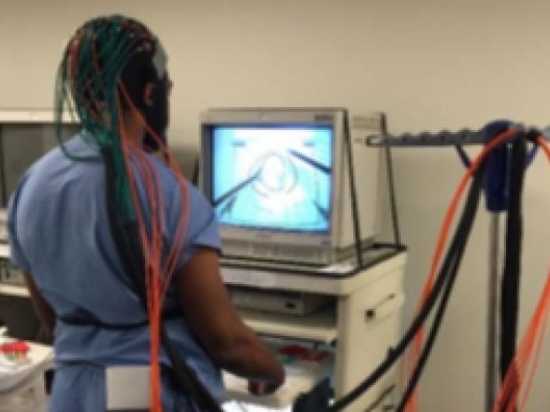
Researchers at Rensselaer Polytechnic Institute envision a day when surgeons will benefit from personalized training, rather than sheer practice repetition, thanks to novel neuroimaging and artificial intelligence methodologies. Under this method, surgeons would complete technical tasks while images of their brain activity reveal how well they have mastered critical skills.
With the support of a $2.2 million grant from the U.S. Army Medical Research and Development Command of the U.S. Department of Defense, received through a Medical Technology Enterprise Consortium award, an interdisciplinary team of researchers at Rensselaer and the University at Buffalo will combine neuroimaging, neuromodulation, and artificial intelligence to better understand and measure surgical skill acquisition — and then determine if that mastery can be accelerated.
“Being able to improve technical skills and certify surgeons based on quantitative metrics is an absolute necessity for a safer surgical environment,” said Suvranu De, the principal investigator on the grant and co-director of the Center for Modeling, Simulation, and Imaging in Medicine (CeMSIM) at Rensselaer. “We need to move toward more objective metrics of skill assessment and certification.”
Surgeons in the United States are currently certified through the Fundamentals of Laparoscopic Surgery and Fundamentals of Robotic Surgery.
These training and testing programs measure skill based on how quickly a surgeon can complete simulated surgical tasks and how many errors they make. De believes this process could be improved if certifiers had a more objective, quantitative measurement of whether a surgeon’s performance reflected a deep level of mastery.
To that end, the research team will use neuroimaging to track where activity is happening within the brain while surgeons are completing technical tasks. Researchers will analyze that data using a collection of deep learning algorithms — known as a deep neural network — to assess and quantify each individual’s level of learning and skill.
“One of the aspects of the study is to better understand how the brain works and how the brain acquires knowledge,” said Xavier Intes, a professor of biomedical engineering and co-director of CeMSIM. “This will be momentous not only for training, but if you have, for instance, a new robotic surgery tool, we can see how the surgeon responds to the new ergonomics or new information feedback, and it can be refined.”
In the second stage of this project, the team will research whether or not neuromodulation can affect neural activity to facilitate learning. In order to do this, researchers will observe the brain activity of medical students who are performing technical tasks while neural stimulation is applied through external electrodes.
This potentially groundbreaking interdisciplinary research is a prime example of The New Polytechnic, the collaborative and innovative spirit with which research at Rensselaer is carried out. For instance, De’s expertise in medical simulation, surgical skill assessment, and patient safety are critically supported by Intes’ expertise in bioimaging and artificial intelligence deep neural networks.
Joining the Rensselaer team are Professors Pingkun Yan and Uwe Kruger from the Department of Biomedical Engineering, and Rahul, a senior research scientist in CeMSIM. The Rensselaer team of engineers is complemented by a team of human factors experts, neuroscientists, and surgeons at the University at Buffalo’s Jacobs School of Medicine and Biomedical Sciences, including Dr. Steven Schwaitzberg, chair of the Department of Surgery, Lora Cavuoto, an associate professor of industrial and systems engineering, and Anirban Dutta, a research associate professor of biomedical engineering and surgery.
“This is a one-of-a-kind project,” De said. “It’s really at the bleeding edge of research at the interface of neuroimaging, deep learning, and surgical skill assessment.”
Sign up for the QuackTrack.org newsletter below!


