


This tool can individually identify signs of neurodegeneration before symptoms appear, which could pave the way for more effective and personalized medicine.
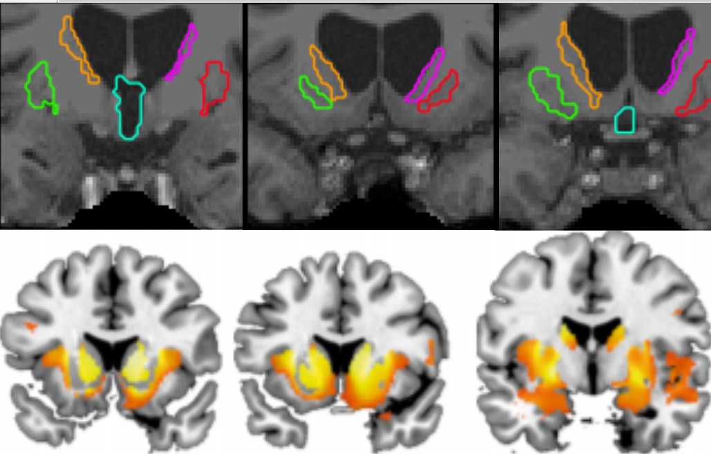
The MRI is the most sensitive and reliable technique for diagnosis and monitoring of neurodegenerative diseases. Through the images obtained we can make a complete anatomical analysis of the brain and detect atrophy in specific regions. However, the inspection of the images is usually visual, which means that certain signs of neurodegeneration, invisible to the human eye, are not detected until the damage is already advanced.
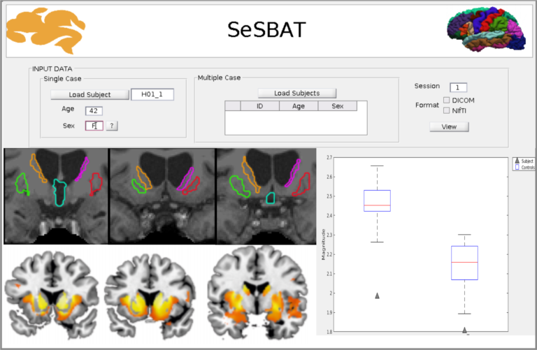
Dra. Estela Càmara from the Bellvitge Biomedical Research Institute ( IDIBELL ) and the Faculty of Psychology and the Institute of Neurosciences of the University of Barcelona ( UBNeuro ), together with Alicia Palomar, have designed a computer program that allows analysis quantitative resonance imaging in an easy, fast, and adaptable way to any brain involvement. This program, called SeSBAT (Single Subject Brain Analysis Toolbox), integrates different image analysis protocols that can measure the volume of specific regions of the brain or perform a general scan of the brain, being able to detect small changes, invisible to the eye, but which could predict the risk of developing a degenerative disease.
SeSBAT opens the door to the identification of new biomarkers that would help improve the early detection and monitoring of neurodegenerative diseases. Being a quantitative analysis it is able to identify anatomical changes in the brain that take place before the onset of symptoms .
With this type of integrated analysis, data can be extracted from a single individual, avoiding the interindividual variability of working with groups. This allows for individualized monitoring of a patient over time, which would be very useful in determining the efficacy of certain drugs in clinical trials, or monitoring people at risk of developing a neurodegenerative disease.
The team of Dra. Camera has tested the accuracy of this new tool on MRI images of patients with Huntington’s neurodegenerative disease. The results, published in the journal Frontiers in Systems Neuroscience , show how this tool is able to identify those patients who are in early stages of the disease, and even those who have not yet shown the first symptoms.
“It is a very versatile program that could be adapted and customized to each type of neurodegenerative disease or project”
Dra. Camera claims that this is a very versatile program that could be adapted and customized to each type of neurodegenerative disease or project. The Chamber group opens the door to new collaborations with groups interested in applying this technology to their projects. Similarly, a tool like this could easily be applied to routine clinical practice, allowing for much deeper analysis of resonance imaging and more accurate management of patients with neurodegenerative diseases.
Sign up for the QuackTrack.org newsletter below!